Pterygium Surgery
Pterygium Surgery
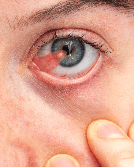
A Pterygium is an outgrowth of the conjunctiva, or mucous membrane, that covers the white part of your eye on the cornea. The cornea is the clear front covering of the eye. Generally, this benign or non-cancerous growth is usually shaped like a wing. Usually, Pterygium isn’t a problem or requires treatment. Also, if it’s affecting your vision. Therefore, It can be a problem and you should have Pterygium Surgery.
What is the reason of this?
Particularly, the exact cause of pterygium is unknown. One explanation is that too much exposure to ultraviolet (UV) light can lead to these growths. However, it is more common in people who live in hot climates and spend a lot of time outdoors in sunny or windy environments. People whose eyes are regularly expose to certain elements are at higher risk of developing this condition.
These elements includes:
- Pollen.
- Annual.
- Smoking.
- Wind.
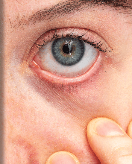
Pterygium Before and After


Slide the bar left end right
What are the symptoms of Pterygium?
Especially, a Pterygium does not always cause symptoms. When it does, the symptoms are usually mild. Common symptoms include redness, blurred vision, and eye irritation. Also, you may feel a burning sensation or itching. If the pterygium grows large enough to cover your cornea, it can affect your vision. Thick or larger Pterygium can also make you feel like you have a foreign body in your eye. As a result, you may not be able to continue wearing contact lenses when you have Pterygium due to discomfort.
How serious is Pterygium?
Pterygium can cause serious scarring of your cornea, but this is rare. Since scarring on the cornea can cause vision loss, it needs to be treated. For minor cases, treatment usually includes eye drops or ointments to treat the inflammation. In more severe cases, it needs to be treated and your Eye Pterygium may need to be surgically remove.
How is it diagnosed?
Diagnosing a Pterygium is simple. Your eye doctor can diagnose this condition based on a physical examination using a Bio-Microscope. It allows your doctor to see by magnifying your eye with the help of Bio-Microscope.
If your doctor needs to do additional tests, they may include:
Visual acuity test:
Especially, this test involves reading letters on an eye chart. In this way, it is determine how much your pterygium affects your vision.
Corneal topographs:
This medical mapping technique is using for measure curvature changes in your cornea.
Digital photography with Bio-Microscope:
This procedure involves taking pictures to monitor the growth rate of the Pterygium.
How is Pterygium treated?
Usually, a pterygium does not require any treatment unless it obstructs your vision or causes severe discomfort. Your eye doctor may want to check your eyes occasionally to see if the enlargement is causing vision problems.
Is it possible to treat with medication?
Especially, if the pterygium is causing a lot of irritation or redness, your doctor may prescribe eye drops or eye ointments containing corticosteroids to reduce inflammation.
Ptergium Surgery
If eye drops or ointments do not provide relief. Your doctor may recommend surgery to remove the pterygium. Surgery is also apply when the pterygium causes vision loss or a condition called astigmatism. Which can cause blurry vision. If you want the pterygium to be remove for cosmetic reasons, you can also discuss surgical procedures with your doctor.
There are several risks associate with these operations. In some cases, a Pterygium may return after surgical removal. After surgery, your eye may be dry and irritate. Your doctor may prescribe medications to provide relief and reduce the risk of pterygium regrowth.
How can I prevent Pterygium?
If possible, avoid exposure to environmental factors that can cause pterygium. Especially, you can help prevent the development of Pterygium by wearing sunglasses or a hat to protect your eyes from sunlight, wind and dust. Your sunglasses should also provide protection from the sun’s ultraviolet (UV) rays. If you already have Pterygium, limiting your exposure to the following can slow its growth:
- Wind.
- Dust.
- Pollen.
- Smoking.
- Sunlight.
By avoiding these conditions, you can also help prevent your previously remove pterygium from returning with surgery.
Pterygium overview
Pterygium surgery is a procedure to remove non-cancerous conjunctival growths (pterygia).
The conjunctiva is the transparent tissue that covers the white part of the eye and the inside of the eyelids. Some cases of Pterygium produce few or no symptoms. An overgrowth of conjunctival tissue can cover the cornea and affect your vision.
Pterygium pre-operative procedures:
Particularly, pterygium surgery is a minimally invasive surgery. It usually takes no more than 30 to 45 minutes. Your doctor will likely provide you with general guidelines to prepare for your pterygium surgery.
You may need to not fast or just eat a light meal before the surgery. Also, if you wear contact lenses, you may be ask not to use them for at least 24 hours before the procedure.
General anesthesia is not used in pterygium surgery. Your pterygium eye is anesthetized with local anesthesia drops. If necessary, your doctor can give you a sedative pill. Therefore, since you cannot drive by yourself, doctors will ask you to arrange transportation after surgery.
Pterygium Postoperative:
At the end of the surgery, your doctor will apply an eye patch or eye pad to ensure your comfort and prevent infection. It is very important that you do not rub your eyes after the procedure to prevent the adhered tissue from being dislodged.
Our doctor will give you aftercare instructions, including cleaning procedures, antibiotics and scheduling a checkup.
It may take a few weeks to several months for your eye to heal completely with no signs of redness or discomfort. However, this may also depend on the type of technique use during the surgery.
Post-operative temporary complications:
As with any surgical procedure, there are risks. It is normal to experience some discomfort and redness after pterygium surgery. It’s also common to notice some cloudiness during healing.
However, please do not hesitate to visit your doctor if you begin to have difficulty seeing, experience a complete loss of vision, or notice that the pterygium has regrowth.
Pinguecula
What is a pinguecula?
Especially, Pinguecula is a benign or non-cancerous growth that develops in your eye. These growths, when more than one, are called pingueculae. These growths occur on the conjunctiva, the thin layer of tissue that covers the white part of your eye.
Pinguecula can be appear at any age, but is mostly found in middle-aged and older people. These growths rarely need to be remove and in most cases no treatment is require.
What does the pinguecula look like?
The pinguecula is yellowish in color and typically has a triangular shape.
It’s a small patch that grows close to your cornea. The cornea is the transparent layer that stretches over your pupil and iris. The iris is the colored part of your eye.
Pinguecula are more common on the side of the cornea near your nose, but they can also grow on the other side near your cornea.
Especially, some Pinguekula can grow, but this happens at a very slow rate and is rare.
What causes pinguecula?
A pinguecula occurs when the tissue in your conjunctiva changes and forms a small lump. Some of these bumps contain fat, calcium, or both. The reason for this change is not fully understand. However, it is link to frequent exposure to sunlight, dust or wind. Pingueculae also tend to become more common as people age.
What are the symptoms of pinguecula?
Pinguecula can make your eye feel irritated or dry. It can also make you feel like you have something in your eye, such as sand or other coarse particles. The affected eye may also itch or become red and inflamed. These symptoms caused by pinguecula can be mild or severe.
Your eye doctor should be able to diagnose this condition based on the appearance and location of the Pinguecula.
Pinguecula vs. Pterygium
More importantly, Pinguecula and Pterygium are types of growths that can form in your eye. These two conditions share a few similarities, but there are also notable differences between them.
Pinguecula and Pterygium are both benign and grow near the cornea. Both are link to exposure to the sun, wind, and other harsh elements.
However, it does not appear like Pterygium. Especially, the pterygium has a tan appearance and is round, oval, or elongate. Pterygium is more likely to grow on the cornea than Pinguecula. A Pinguecula growing on the cornea can be popularly confuse with a Pterygium.
How is pinguecula treated?
You usually do not need any treatment for Pinguecula unless it is causing discomfort. If your eye hurts, your doctor may give you eye ointment or eye drops to relieve redness and irritation.
If its appearance bothers you, you can talk to your doctor about surgical removal of the Pinguecula. In some cases, your Eye Surgeon may need to surgically remove the Pinguecula so that it does not grow any further.
The pinguecula may need to be surgically removed in the following cases:
- In cases where your cornea grows on and affects vision.
- When it causes extreme discomfort in contact lens wearers.
Especially, in the treatment of pinguecula, it may be require to be remove by the Eye Surgeon in cases where continuous. Also, severe inflammation and side or redness continues even after applying eye drops or ointment.
How is its development in the long run?
Pinguecula usually does not cause any problems. The pinguecula may grow back later, but surgery does not typically lead to complications. Your doctor may give you medicine to help prevent it.
Can you prevent Pinguecula from developing?
You are more likely to develop Pinguecula if you spend a lot of time outside because of your work or daily life. However, you can help prevent these growths by wearing sunglasses when outside. Therefore, you should wear sunglasses with a coating that blocks the sun’s ultraviolet A (UVA) and ultraviolet B (UVB) rays. Also, sunglasses helps protect your eyes from wind and other external factors such as sand.
Moisturizing your eyes with artificial tears can also help prevent pinguecula. You should also wear safety glasses when working in a dry and dusty environment.
Useful Links
Strabismus Crossed Eye Squint Treatments, Diabetic Retinopathy Disease. Macular Degeneration Treatments. Glaucoma Disease and Treatments. Retinitis Pigmentosa Disease and Treatments. Periodic Eye Examinations.
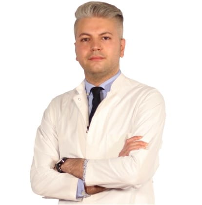
Your Expert Eye Surgeon

Education Information
He completed primary, secondary and high school in Baku. He graduated from Azerbaijan Medical Faculty in 2010. He completed his ophthalmology residency in Baku National Ophthalmology Center Azerbaijan . He was entitled to receive the Presidential Scholarship of the Republic of Azerbaijan in 2014, and completed the equivalence of specialization in Turkey in the Department of Ophthalmology, Faculty of Medicine, Selcuk University in 2014-2018, and worked as a research assistant and ophthalmologist at the university until 2018. He worked as an Ophthalmologist at Private LIV Hospital Nation between 2019-2022.Work experience:
- Selcuk University Faculty of Medicine
- Private LIV Hospital Ulus
Certificates, Memberships, Scientific Research:
- Peroperative developing choroidal detachment and its management.
- Surgical Approach in Posterior Polar Cataract.
- Iatrogenic retinal artery occlusion caused by cosmetic facial autologous fat filler injections.
- Effect of Smoking on Ocular Surface and Corneal Nerves.
- Lupus choroidopathy in a patient with discoid lupus erythematosus.
- Endophthalmitis and its treatment with early parsplanavitrectomy.
- Turkish Ophthalmology Association.
Specialized Treatments and Surgeries:
- Retractive Laser Eye Surgery: iLASIK (Femto Lasik), LASIK, LASEK, Trans Epithelial PRK (NO TOUCH) and SMILE
- Cataract Surgery (Smart Lens Surgery)
- Keratoconus Treatments – Cross-Linking Surgeries
- Pterygium Surgery
- Dry Eye Disease and Treatments
- Strabismus Surgery
- Glaucoma- Glaucoma Eye Pressure Treatments
- neuro ophthalmology
- Retinal Diseases Treatments
- Oculoplasty
- Uveitis Diseases
- Ectropion and entropion surgery – Eyelid deformity treatments
- Enucleation and Evisceration Prosthetic Eye Surgery
Foreign language:
- English
- German
- Russian
- Azerbaijani
- Turkish
MEDICAL UNIVERSITY of Selcuk
12 Years of Experience
>8.000 Surgery
Definitely avoid low-cost Eye Surgery
You may think that a cheap eye surgery is right for you. This might be fine if you're buying a cheap TV, but it's not worth the gamble with your eyesight. But as you know, having cheap eye surgery means sacrificing technology, physician quality, medical care and sterile conditions, and most importantly, taking risks. The issues that fall on a patient who wants to have cataract surgery and should pay the most attention; The hospital with the latest technology in cataract surgery and imaging devices, a sterile environment, and an experienced doctor and clinical team should be selected. We would like to remind all our patients that they only have two eyes and that the most important and most sensitive sense organ is their eyes.
None of these things are more important than your eye health, and we do not compromise on quality and cutting-edge technology. We offer you our prices in a very understandable, fair and affordable way.
There are no hidden costs in our pricing. We make eye surgery affordable for you.


